Onychodermal Band: The Distal Seal Between Nail Plate and Nail Bed
Author: Radina Ignatova, Professional Nail Expert & International Nail Educator | Last Updated: May 2026
Quick Answer: The onychodermal band is the narrow transverse band of specialised epithelium at the distal end of the nail bed — the last point of firm adhesion between the nail plate and the nail bed before the plate separates into the free edge. It is sometimes visible through the nail plate as a faint pale or pinkish-white band just proximal to where the white of the free edge begins. It works in close functional relationship with the hyponychium — together they form the distal seal of the nail unit.
Contents
Anatomy of the Onychodermal Band
The onychodermal band — from the Greek onyx (nail) and derma (skin) — is a narrow band of specialised epithelial tissue located at the very distal end of the nail bed. It marks the boundary between where the nail plate is firmly adhered to the nail bed and where it begins to separate into the free edge. This transition is not abrupt — the onychodermal band is the zone where adhesion progressively reduces before separation is complete.
The band runs transversely across the full width of the nail — from one lateral groove to the other — at the point where the vascular, pink nail bed ends and the nail plate begins to lift. It is a fixed anatomical structure, though its exact width and prominence vary between individuals.
The onychodermal band sits just proximal to the hyponychium. The hyponychium begins at the distal side of the onychodermal band — where the nail plate has fully separated from the nail bed and the free edge begins. The two structures work together as the distal seal of the nail unit: the onychodermal band provides the last adhesive anchor, and the hyponychium provides the physical barrier beyond it.
Visibility Through the Nail Plate
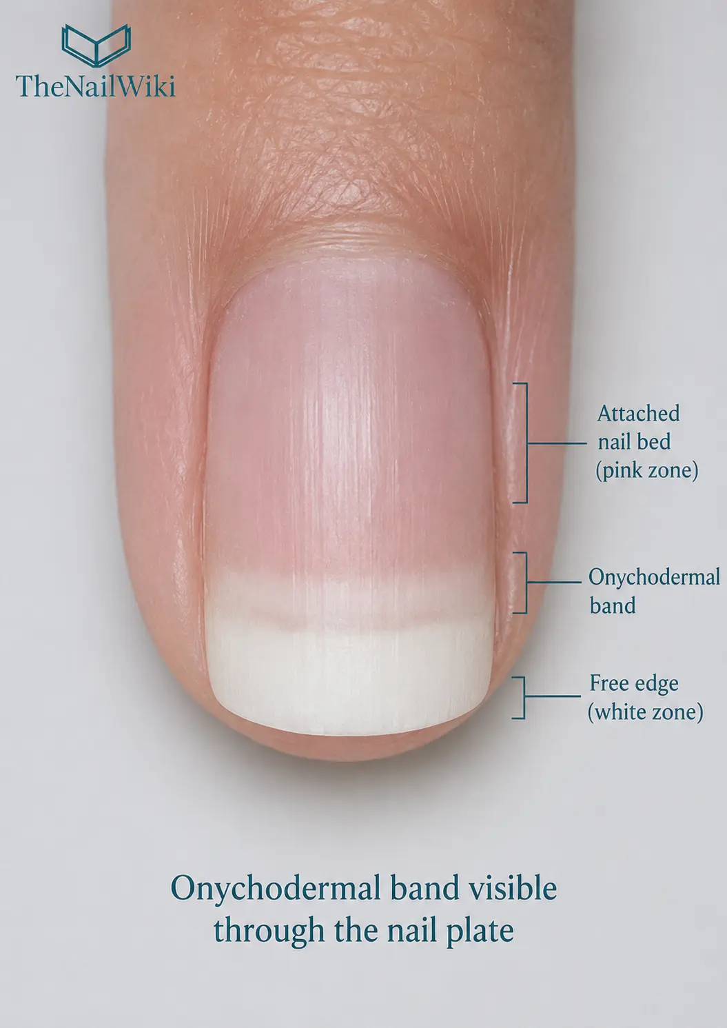
The onychodermal band is sometimes visible through the translucent nail plate as a faint transverse band — slightly paler, pinker, or whitish in appearance — running across the width of the nail just inside the proximal boundary of the white free edge. It is not always visible, and its prominence varies between clients and between digits on the same hand.
When visible, it creates a subtle but distinct transition zone between the fully pink attached nail plate — where the nail bed transmits its colour through the translucent plate — and the white free edge — where no nail bed underlies the plate and the plate appears opaque. The onychodermal band sits within this transition, appearing slightly different from both zones because the adhesion here is reducing but not yet absent.
The visibility of the onychodermal band is not clinically significant in itself — its absence does not indicate a problem and its presence does not require intervention. It is simply a feature of the distal nail anatomy that becomes relevant as a reference point when assessing the distal nail unit and when identifying where onycholysis begins.
The three distal zones visible through the nail plate
- Pink zone — the fully adhered nail plate over the nail bed. The blood-rich nail bed transmits its colour through the translucent plate
- Onychodermal band — a faint pale or pinkish-white transverse band where adhesion is reducing. Not always visible
- White free edge — the nail plate with no nail bed beneath it. No colour transmits — the plate appears white or opaque
© TheNailWiki
Function — Distal Adhesion and Sealing
The onychodermal band’s primary function is to provide the final zone of firm adhesion between the nail plate and the nail bed at the distal end of the nail. The nail plate adheres to the nail bed across its full length — but this adhesion does not terminate sharply at a single line. The onychodermal band is the zone where that adhesion gradually reduces to zero as the plate transitions into the free edge.
This gradual transition is structurally important. A sudden termination of adhesion at a hard boundary would create a concentrated stress point — the plate would be fully anchored on one side of the line and completely free on the other, generating a shear stress every time the nail flexes. The onychodermal band smooths this transition, distributing the adhesion reduction across a small zone rather than concentrating it at a single point.
Together with the hyponychium, the onychodermal band forms the distal barrier of the nail unit. The onychodermal band anchors the plate. The hyponychium seals the gap created where the plate separates. As long as both are intact, the subungual space — the space between the nail plate and the nail bed — is effectively closed at its distal end.
The Onychodermal Band and Onycholysis
Onycholysis — the separation of the nail plate from the nail bed — almost always begins at or distal to the onychodermal band. When the onychodermal band is disrupted, the nail plate separates from the last point of firm adhesion and a pocket begins to form between the plate and the nail bed. The hyponychium, which seals the distal side of this boundary, can no longer maintain its contact with the underside of the now-separated plate.
The proximal boundary of onycholysis — how far back from the free edge the separation extends — is typically assessed by observing where the white or discoloured zone ends and the pink attached nail begins. This boundary corresponds to where the onychodermal band previously sat. As onycholysis progresses, this boundary moves proximally — further from the free edge — indicating that the separation is extending beyond the onychodermal band zone into the more firmly adhered nail bed.
For nail professionals, the position of the onycholysis boundary relative to the normal onychodermal band position is a useful clinical reference. Separation that extends only slightly beyond the onychodermal band — a small white arc at the free edge — may be early or mild. Separation that extends well into the mid-plate area represents a more significant disruption of the nail bed adhesion. See: Onycholysis →
Causes of onychodermal band disruption
- Trauma — a direct blow, compression, or forcible impact at the free edge
- Repeated cleaning beneath the nail — pushing instruments under the plate disrupts the band progressively
- Chemical exposure — harsh cleaning products, solvents, sulphur-containing mineral pools
- Aggressive filing at the free edge — directing the file beneath the plate rather than across its tip
- Systemic conditions — thyroid disease, nail psoriasis, certain medications
- Contact sensitisation — uncured nail product contacting the distal nail unit
Relevance to Professional Nail Services
As a reference point in client assessment
The onychodermal band is a useful reference in the pre-service nail assessment. When visible, it marks the expected boundary between the attached nail plate and the free edge. Any white or discoloured zone that extends proximal to the onychodermal band indicates onycholysis — the plate is separating beyond its normal distal boundary. Recognising this distinction is part of professional nail assessment before any service or product application.
Free edge sealing and the onychodermal band
In gel polish and regular polish services, sealing the free edge — applying product across the tip of the nail — protects the onychodermal band and hyponychium from moisture and solvent ingress. Product that enters beneath the plate at the distal end directly contacts the onychodermal band zone and over time contributes to its disruption. See: Free Edge →
Product application at the distal nail
When applying gel or enhancement product to the nail plate, the onychodermal band is the distal anatomical boundary of the nail bed. Product should not be forced beneath the plate distal to this boundary. Any product that contacts the hyponychium or the onychodermal band zone beneath the plate — rather than being applied to the top surface — risks sensitisation and contributes to distal seal disruption. See: Hyponychium →
Frequently Asked Questions
What is the onychodermal band?
The onychodermal band is the narrow transverse band of specialised tissue at the distal end of the nail bed — the last zone of firm adhesion between the nail plate and the nail bed before the plate separates into the free edge. It is sometimes visible as a faint pale band just inside the boundary of the white free edge.
What is the difference between the onychodermal band and the hyponychium?
They are adjacent structures with different roles. The onychodermal band is the last zone of adhesion between the nail plate and nail bed — it is part of the nail bed. The hyponychium is the skin seal at the distal end of the nail unit, sitting beyond the onychodermal band where the nail plate separates. The onychodermal band anchors the plate distally; the hyponychium seals the gap beyond that anchor.
Is it normal to see the onychodermal band?
Yes — it is visible in some clients and not others, and varies between digits. Its visibility is not clinically significant. It appears as a faint pale or pinkish-white transverse band just proximal to the white free edge, and represents the normal transition zone between attached and free nail plate.
How does the onychodermal band relate to onycholysis?
Onycholysis begins when the onychodermal band is disrupted — the nail plate separates from its last distal anchor point and a pocket forms beneath the plate. The proximal boundary of onycholysis corresponds to how far the separation has extended beyond the normal onychodermal band position. The further proximal this boundary, the more extensive the separation.
Can the onychodermal band heal after onycholysis?
Yes — once the cause of onycholysis is removed and the nail bed is kept clean and dry, the nail plate can reattach to the nail bed as new growth advances. The onychodermal band re-establishes at the distal end of the reattaching plate. Recovery requires removing the cause, keeping the separated area clean, and allowing new nail plate growth to advance and re-adhere to the nail bed — which takes time proportional to how far the separation extended.
Professional training in nail anatomy
Nail anatomy, client assessment, and professional nail services are taught as part of structured courses at Artistic Touch Nail Training Academy.
Related Library Pages
Nail Anatomy
Conditions
Professional Disclaimer
The information on this page is provided for educational purposes and is intended to support the professional knowledge of nail technicians and nail educators. It does not constitute medical advice. Any persistent nail or skin changes should be assessed by a qualified medical professional.
About the Author
Radina Ignatova
Professional Nail Expert | International Nail Educator
Radina Ignatova is a Professional Nail Expert since 2014, International Nail Educator, and Founder of TheNailWiki and Artistic Touch Nail Training Academy. She specialises in Russian Manicure, dual form systems, polygel, advanced e-file techniques, and nail safety protocols, and continues to work actively in salon practice ensuring that all education reflects real client scenarios and current industry standards.
Her teaching philosophy is built on honest education — showing real salon challenges, real mistakes, and real performance testing rather than presenting only perfect demonstrations. This is how genuine technical competence is developed and how nail professionals become truly confident and capable.
Read full bio →© 2026 TheNailWiki — an independent nail education resource. Content is safety-led and professionally informed.