
Normal White Free Edge or Early Onycholysis? Using the Onychodermal Band to Tell the Difference
Quick Answer: Is a White Tip Normal or Is the Nail Lifting?
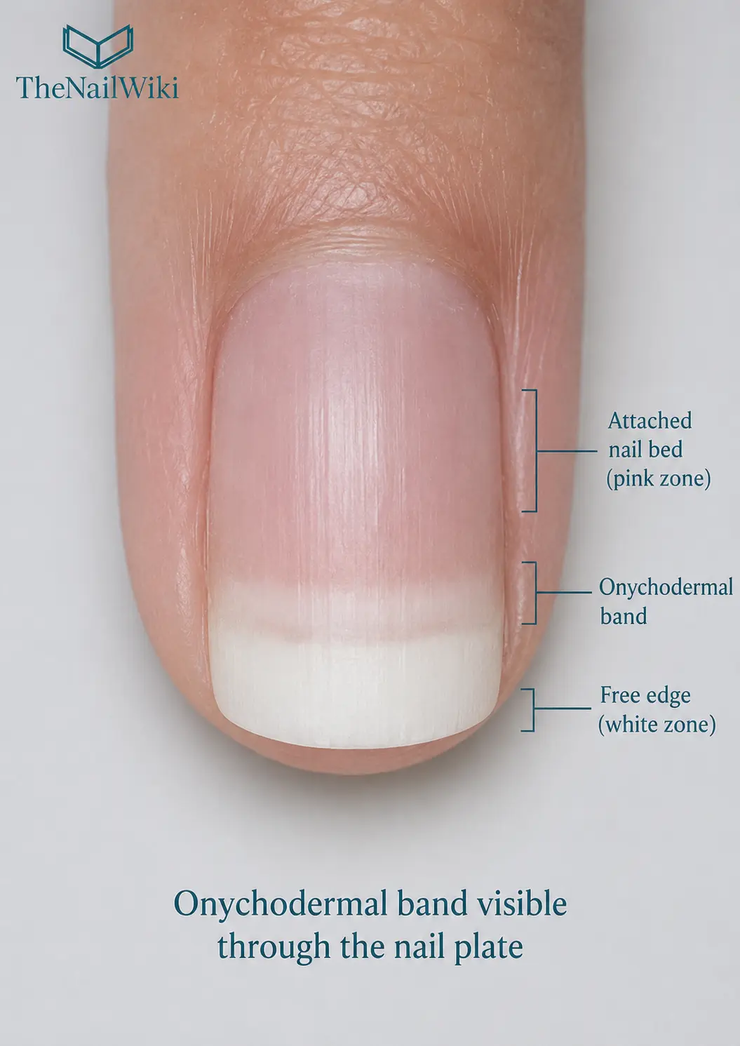
A white free edge is normal, and a white or discoloured zone that extends back beyond the normal boundary of attachment is not. The onychodermal band marks the last point of firm adhesion between the nail plate and the nail bed. Where it can be seen, it shows where the pink attached nail should end and the white free edge should begin. A white or discoloured area proximal to that line indicates onycholysis, meaning that the plate is separating beyond its normal distal boundary.
Why it matters: early separation is easy to miss under polish or product, and assessing it before a service prevents sealing a problem beneath a new coating.
The Three Zones at the Tip of the Nail
Looking at the distal nail through the translucent plate, three zones can usually be distinguished. They are the reference for everything that follows.
- The pink zone. The plate is firmly attached to the nail bed, and the blood-rich bed shows its colour through the plate.
- The onychodermal band. A faint, slightly paler or pinkish-white transverse line where adhesion is reducing. It is not always visible and its prominence varies between clients and between fingers.
- The white free edge. No nail bed lies beneath the plate here, so no colour is transmitted and the plate appears white or opaque.
The visibility of the band is not significant in itself. Its absence does not indicate a problem, and its presence needs no intervention. It is simply a landmark. Beyond it, the hyponychium forms the seal where the plate has fully separated from the bed.
What Normal Looks Like
A normal tip has a clear change from pink to white, with the white zone beginning at a consistent line across the width of the nail. The boundary is usually smooth and follows the curve of the fingertip. The nail may look a little different on each finger, because the length of the free edge and the visibility of the band vary between digits.
It helps to learn what is normal for each client, since the position of the boundary is individual. A client's own earlier appointments are a better reference than a general rule.
What Early Separation Looks Like
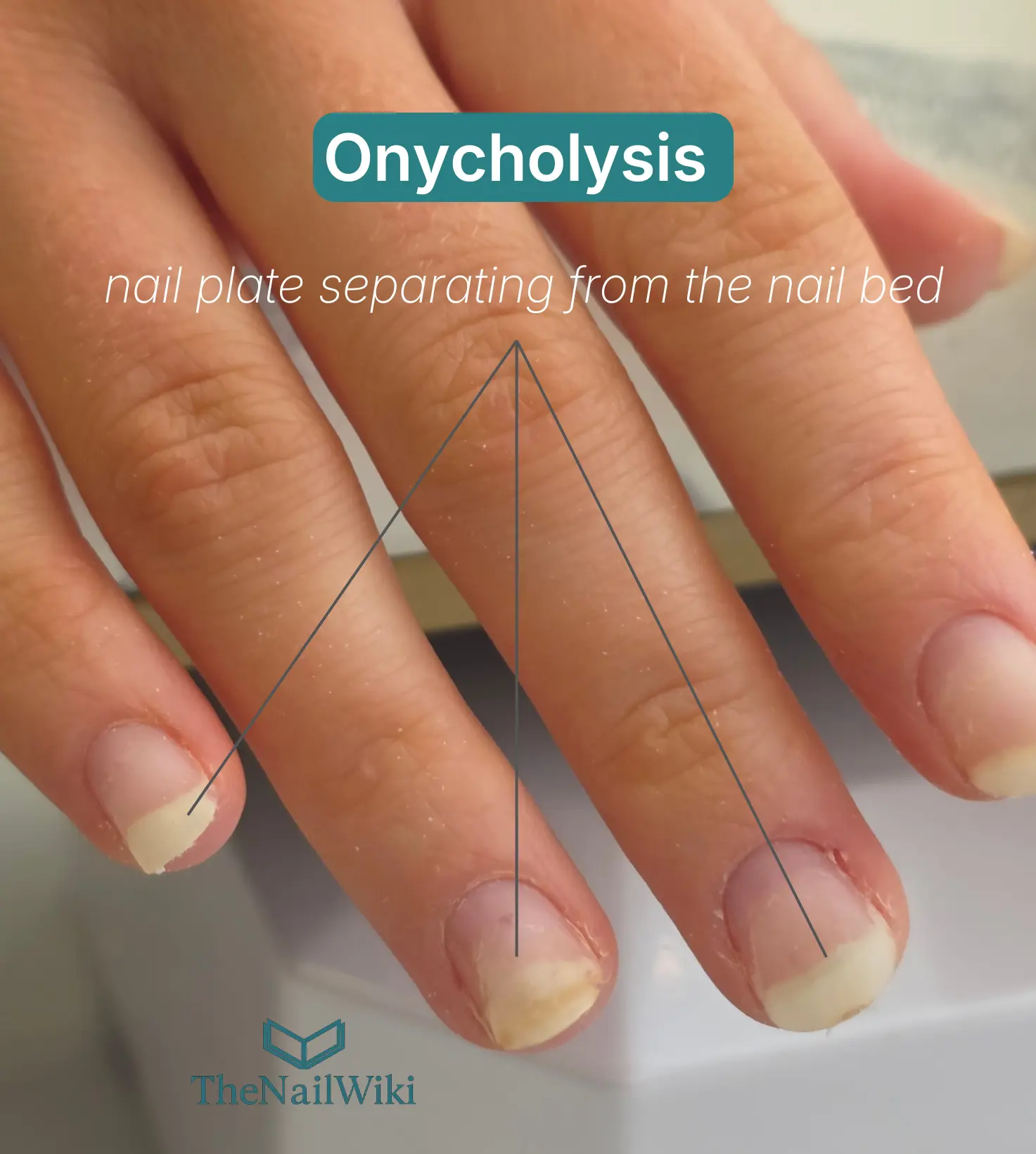
Onycholysis almost always begins at or beyond the onychodermal band. As it progresses, the boundary between the pale area and the pink attached nail moves proximally, away from the tip. The position of that boundary, compared with where the band would normally sit, gives a practical indication of how far the separation has gone.
- A small white arc at the free edge may indicate early or mild separation.
- A boundary that has moved well into the mid-plate represents a more significant disruption of attachment.
- A change of colour in the pale zone, such as yellow or green, suggests that the space beneath the plate may have become contaminated.
Separation under a coating is not always visible until the product is removed. A hollow sound when the nail is gently tapped, or sensitivity at the free edge, can be early clues. The article Why Do Nails Lift? explains the causes in detail.
How the Band Gets Disrupted
The Library lists several causes of disruption, and many of them are things a client does or has done to them rather than a feature of the nail itself.
- Trauma, such as a blow or compression at the free edge
- Repeated cleaning beneath the nail with a tool
- Exposure to harsh cleaning products, solvents or sulphur-containing mineral pools
- Filing at the free edge in a way that directs the file beneath the plate rather than across its tip
- Systemic conditions such as thyroid disease or nail psoriasis, and certain medications
- Contact sensitisation from uncured product reaching the distal nail unit
Because some causes are medical, a pattern that does not respond to changes in habits or technique is a reason to suggest assessment. See nail psoriasis and the guide to contraindications in nail services.
Applying Product at the Distal Nail
The band is also the distal boundary of the nail bed, which gives a practical rule for product. Gel or enhancement product belongs on the top surface of the plate, and it should not be forced beneath the plate beyond the band. Product that contacts the hyponychium or the band zone beneath the plate risks sensitisation and contributes to disrupting the distal seal.
Sealing the free edge, which means applying product across the tip, protects the band and the hyponychium from moisture and solvents entering beneath the plate. Product that gets beneath the plate at the tip contacts the band zone and, over time, contributes to its disruption. See free edge and the article Common Free Edge Problems.
A Simple Response Ladder
Match the response to what you see
- Normal boundary, healthy tissue: proceed as usual and keep a note of the client's normal boundary.
- Small arc that is new or changing: ask about habits, cleaning and products; avoid probing beneath the plate; reassess at the next appointment.
- Separation extending into the plate, discolouration or debris beneath: do not seal it beneath a coating. Remove product, keep the area clean and dry, and refer where appropriate.
- Pain, swelling, heat or signs of infection: stop the service and refer the client to a medical professional.
Can the Band Recover?
Yes. Once the cause is removed and the area is kept clean and dry, the plate can reattach to the nail bed as new growth advances, and the band re-establishes at the distal end of the reattaching plate. Recovery takes time in proportion to how far the separation extended, because new plate must grow forward over the bed.
Frequently Asked Questions
Is it normal to see the onychodermal band?
Yes, in some clients and on some fingers. Its visibility is not clinically significant, and not seeing it is also normal.
How do I know whether a white tip is lifting?
Compare the boundary of the white zone with where the pink attached nail should end. A white or discoloured zone that extends proximal to the normal position of the band indicates that the plate is separating.
What is the difference between the onychodermal band and the hyponychium?
The band is the last zone of firm adhesion and is part of the nail bed. The hyponychium is the skin seal beyond it, where the plate has separated from the bed.
Can a nail that has lifted reattach?
It can, once the cause is removed and the area is kept clean and dry, as new plate grows forward and adheres to the bed.