Hyponychium: The Seal Beneath the Free Edge
Author: Radina Ignatova, Professional Nail Expert & International Nail Educator | Last Updated: May 2026
Quick Answer: The hyponychium is the thickened epithelial tissue beneath the free edge of the nail plate — the point where the nail separates from the nail bed at the fingertip. It forms a waterproof seal at the distal end of the nail unit, closing off the subungual space from external contamination. At the opposite end of the nail, the eponychium seals the proximal end — together they protect the entire underside of the nail plate from pathogens, moisture, and debris.
Quick Summary
The hyponychium is one of the least discussed structures in the nail unit — yet it is clinically significant and directly relevant to some of the most common problems nail professionals encounter, including onycholysis, green nail syndrome, and lifting at the free edge. It contains its own blood supply and nerve endings — it is living, sensitive tissue, not simply a patch of thickened skin. Disrupting it — through aggressive cleaning beneath the nail, forced pushing, or incorrect product technique — has real consequences for nail health and infection risk.
The hyponychium is also the structure responsible for the characteristic discomfort some clients report when wearing extensions that are too long — it bears the mechanical load transferred from the free edge of the enhancement to the fingertip beneath it.
Contents
- Anatomy of the Hyponychium
- Function — Sealing the Distal Nail Unit
- The Hyponychium and the Onychodermal Band
- Why Cleaning Beneath the Nail Damages the Seal
- Hyponychium Adaptation — How It Changes With Nail Length
- Relevance to Professional Nail Services
- Conditions Affecting the Hyponychium
- Frequently Asked Questions
Anatomy of the Hyponychium
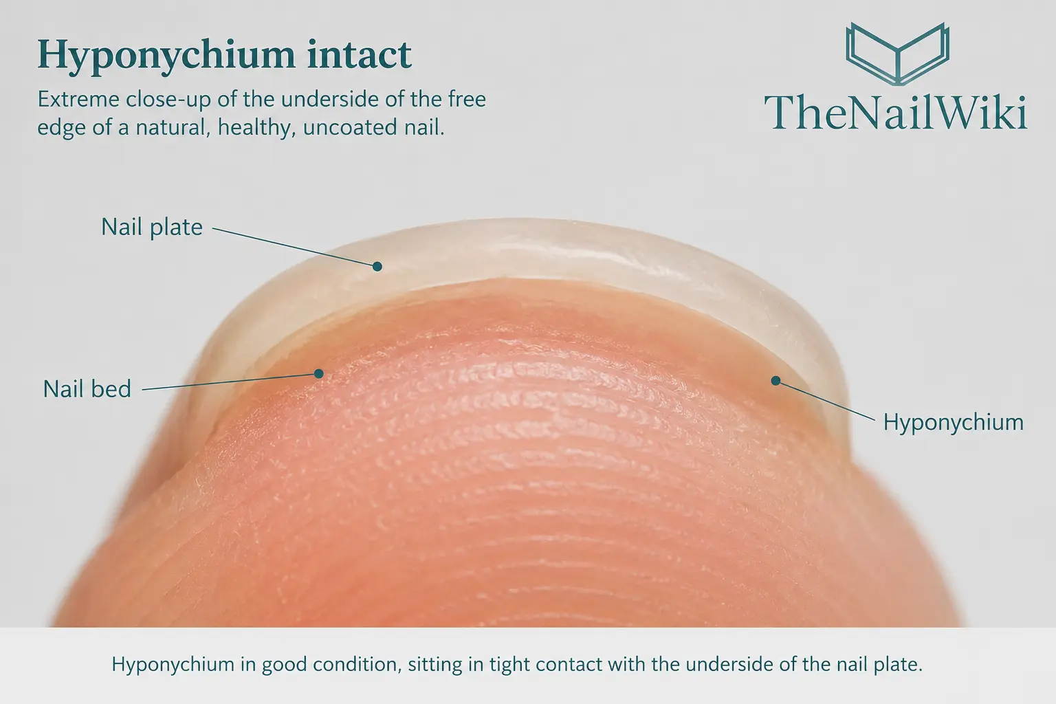
The thickness and health of the hyponychium directly reflects the thickness and health of the nail plate above it. Clients with naturally thin nail plates tend to have a less substantial hyponychium — thinner, more easily disrupted, and more vulnerable to both chemical and mechanical damage. Clients with thick, robust nail plates typically have a correspondingly more substantial hyponychium that is more resistant to disruption. This is also why damage to the nail plate — through over-filing, aggressive services, or repeated mechanical trauma — directly increases the vulnerability of the hyponychium beneath it.
The hyponychium sits in close anatomical relationship with the onychodermal band — a narrow zone of epithelium just proximal to it that marks the last point of firm nail-plate-to-nail-bed adhesion before the plate separates into the free edge. The onychodermal band is often visible through the translucent nail plate as a faint pale or pinkish-white line just proximal to where the white free edge begins.
Key anatomical facts
- The hyponychium is thickened epithelium — not callus and not keratin build-up — it is a normal, specialised tissue structure
- It has its own blood supply and nerve endings — it is living, sensitive, innervated tissue
- It sits at the distal end of the nail unit, at the opposite end from the eponychium
- Its primary function is barrier protection — it closes the subungual space from the external environment
- It adapts to nail length — growing forward beneath longer nails and retracting as nails are shortened
- The transition from hyponychium to free edge is marked by the onychodermal band just proximal to it
Function — Sealing the Distal Nail Unit
The hyponychium’s primary function is to form a physical and biological barrier at the distal end of the nail unit — preventing bacteria, fungi, moisture, and particulate debris from entering the subungual space between the nail plate and the nail bed. The subungual space, if contaminated, becomes a site for infection that is difficult to treat because the nail plate sits over it, creating a closed environment that shields the contamination from both topical treatments and air.
The sealing system of the nail unit works on three sides. The eponychium seals the proximal end. The lateral nail folds seal the sides. The hyponychium seals the distal end. When all three are intact, contaminants cannot easily access the nail bed. When any one is disrupted, the nail bed becomes vulnerable — and the hyponychium, because it is directly adjacent to the open environment of the fingertip, is the most frequently compromised of the three.
The hyponychium also contributes to the mechanical attachment of the nail plate at its distal edge. The nail bed provides the primary adhesion for the nail plate across its length — but the hyponychium’s contact with the underside of the plate at the very tip adds the final point of stabilisation before the plate becomes unsupported at the free edge. When the hyponychium is disrupted and onycholysis begins, the nail plate loses both its distal adhesion and its distal seal simultaneously.
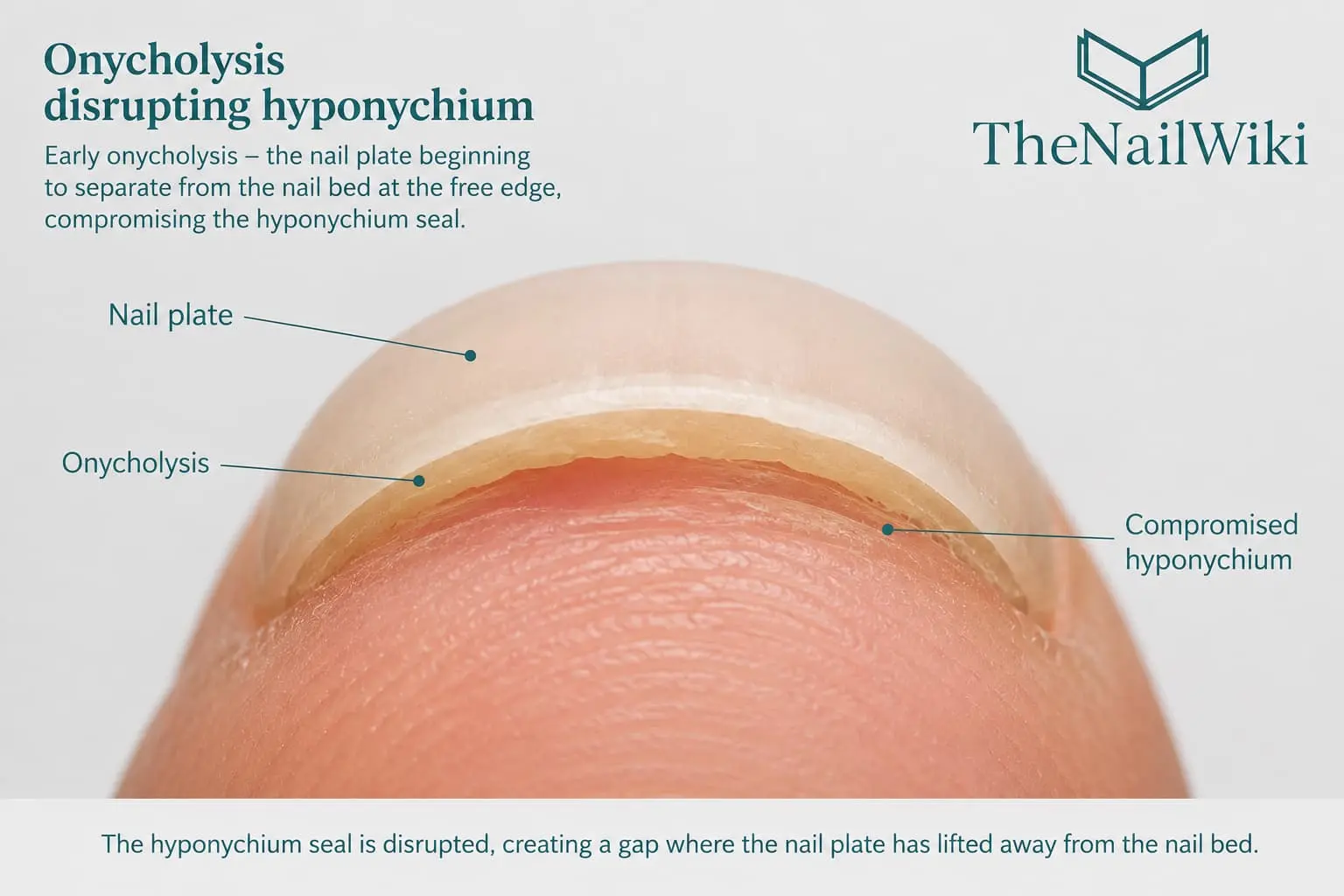
The Hyponychium and the Onychodermal Band
The onychodermal band is a narrow transverse band of epithelium at the distal end of the nail bed — located just proximal to the hyponychium, at the point where the nail plate and nail bed are most firmly adherent before separating. It is visible through the nail plate as a faint whitish or pale pinkish line running across the width of the nail, just inside the boundary of the free edge.
The onychodermal band represents the last point of firm nail-plate-to-nail-bed adhesion. Proximal to it, the nail plate is firmly bonded to the nail bed. Distal to it, the plate separates progressively and the hyponychium takes over the sealing role. The onychodermal band also has its own colour contribution — it is slightly less transparent than the overlying plate, which is why it is sometimes visible as a distinct pale band even in healthy nails.
In onycholysis, the onychodermal band is disrupted — the separation extends proximal to its normal position, and the hyponychium can no longer maintain its seal against the underside of the now-detached plate. The resulting pocket fills with moisture, keratin debris, and, in some cases, bacterial or fungal organisms.
For nail professionals, understanding the onychodermal band and hyponychium as a functional unit — rather than simply as the area where the white of the free edge begins — is important for correctly assessing clients who present with free edge changes, discolouration, or early onycholysis.
Why Cleaning Beneath the Nail Damages the Seal
One of the most common causes of hyponychium disruption is a habit that many clients consider good nail hygiene — cleaning beneath the free edge with a pointed implement to remove debris. This practice is particularly common among clients who feel that the space under the nail collects dirt and that cleaning it out is necessary for cleanliness.
In reality, the opposite is true. The hyponychium forms a natural barrier that keeps the subungual space clean by preventing debris from entering in the first place. When an instrument is pushed beneath the free edge and used to lever or scrape beneath the nail plate, it mechanically separates the hyponychium from the underside of the plate — each time it is done, the seal is disrupted slightly further back. Over time, this progressively enlarges the subungual space, allowing more debris and moisture to accumulate rather than less.
Mechanical disruption is not limited to tools pushed directly beneath the nail. Using the nail plate itself as a lever — for example, peeling an orange, prising open packaging, or using the nail tip to scrape surfaces — applies strong downward and lateral forces to the free edge zone, transmitting stress through the hyponychium and progressively breaking its contact with the underside of the plate. Using a nail brush with bristles that are too coarse to clean beneath the free edge can also mechanically abrade the hyponychium and widen the subungual space over time. These habits are so normalised in daily life that clients rarely connect them to the nail changes they present with.
The progressive disruption of the hyponychium through repeated undercleaning creates exactly the conditions that cause onycholysis — nail plate separation from the nail bed beginning at the free edge. Once separation exists, the moist, warm, enclosed space between the plate and the bed becomes an ideal environment for Pseudomonas colonisation, producing green nail syndrome, or for fungal growth.
What to tell clients about cleaning beneath the nail
The correct approach is to clean the visible underside of the free edge — the edge of the nail plate that is visible looking at the fingertip — without pushing anything beneath the plate itself. The hyponychium is not debris; it is a functional structure. Disturbing it creates the contamination risk the client is trying to prevent. This is one of the most useful pieces of education a nail professional can give a client, and it directly connects cleaning habits to infection risk in a way that is easy to understand.
Safe cleaning beneath the nail
For clients who want to clean beneath the free edge, the correct approach is a soft-bristled brush — used gently across the underside of the free edge without digging in or pushing the bristles under the plate itself. A soft brush removes surface debris from the visible underside without disrupting the hyponychium seal beneath. Any brush with stiff bristles, or any brushing technique that pushes under the plate rather than across the edge, risks the same progressive mechanical disruption as a tool.
Chemical exposure — what breaks the hyponychium seal
The hyponychium seal is more chemically vulnerable than most clients realise. Exposure to certain environments and products can break down the epithelial tissue and disrupt the seal in a single exposure — not requiring the repeated mechanical disruption described above. Sulphur-containing mineral hot pools are a direct example — the sulphur compounds are strongly oxidising and acidic, and prolonged immersion can dissolve the hyponychium seal in the same way that extended bleach exposure does. Household bleach-based cleaning products, harsh detergents, and even extended dishwater exposure all progressively degrade the seal over time.
Clients whose nail plates are already thinned or damaged — whether from previous over-filing, repeated aggressive services, or medical conditions — are significantly more susceptible. A compromised nail plate offers less mechanical protection to the hyponychium beneath it, so chemical disruption occurs faster and with less exposure than in a client with a healthy, full-thickness plate.
Protecting the hyponychium from chemical exposure
- Wear rubber or nitrile gloves for all cleaning tasks involving bleach, detergents, or harsh household products
- Avoid prolonged immersion of the hands in mineral pools, particularly sulphur-based hot pools — if swimming or soaking, keep exposure to a minimum and apply cuticle oil afterwards
- Rinse hands thoroughly after any chemical exposure before applying any nail product or oil
- Clients with already damaged or thinned nail plates should be particularly careful — their hyponychium seal is more vulnerable to disruption from shorter exposure
Cuticle oil and the hyponychium
Cuticle oil is most commonly applied to the proximal nail fold and cuticle area — but the hyponychium is an equally important target. The seal at the distal end of the nail is skin tissue like any other, and it benefits from the same hydration and conditioning that the proximal skin receives. A hyponychium that is well hydrated remains flexible and resilient — it maintains firm contact with the underside of the nail plate more effectively than one that is dry and brittle.
Applied to the hyponychium, cuticle oil also fills the very small gap at the distal end of the nail — providing a thin layer of oil that acts as a barrier between the plate and any debris or moisture trying to enter the subungual space. This is particularly valuable for clients who spend time in water, work with their hands, or are recovering from onycholysis. A missed space when applying cuticle oil is the underside of the free edge and the hyponychium — applying oil here as well as to the cuticle area completes the treatment.
Hyponychium Adaptation — How It Changes With Nail Length
The hyponychium is not static — it adapts to the position of the nail plate over time. As nails grow longer or as nail extensions are worn, the hyponychium gradually migrates forward beneath the nail plate, maintaining its seal at the advancing edge of the free edge. This is a biological adaptation — the tissue follows the structure it is sealing, ensuring that the subungual space remains protected regardless of free edge length.
This adaptation is most visible in clients who have worn long nail extensions for extended periods. When the extensions are finally removed and the natural nail is kept shorter, the hyponychium has grown forward to follow the longer plate — it is now visible as a visible ridge of skin beneath the shorter free edge of the natural nail. This is sometimes alarming to clients who feel that something has grown over or under their nail and needs to be removed.
An overgrown hyponychium of this kind is not pathological. It is normal adaptive tissue that has followed the nail. It should not be cut, filed, trimmed, or forced back — doing so breaks the seal, risks bleeding, and can be significantly painful given the nerve supply in this tissue. As the nails are kept at a shorter length over time — typically over several months — the hyponychium gradually retracts on its own, following the plate back to its new position.
In some cases — particularly in clients with hooked or downward-growing nails — the hyponychium can grow forward and upward onto the nail plate surface itself rather than simply following the plate beneath it. When the nail plate curves downward at the free edge, the hyponychium follows the curve and can begin to adhere to the top surface of the plate at the tip rather than remaining beneath it. This can make the free edge appear to have skin growing over it and can make shortening the nail uncomfortable or difficult. This is not a pathological condition — it is an adaptation to the nail’s growth direction — but it does require careful technique when shaping or filing the free edge. Cutting into this tissue causes pain and bleeding.
Similarly, in clients who bite their nails very short, the hyponychium retracts and thins over time. When these clients begin growing their nails out — or begin wearing extensions — the hyponychium has not yet adapted to the new plate length. The subungual space at the free edge may therefore be more open initially, and these clients can be more susceptible to debris accumulation and infection during the transition period.
Relevance to Professional Nail Services
E-file work near the hyponychium — professional technique and client safety
In professional nail services — particularly when removing the free edge before hard product application, or when cleaning the underside of the free edge in dual form or Sandwich Dual Form services — the e-file works in close proximity to the hyponychium. This requires particular care and attention to one factor that is frequently underestimated: the client must not move during this work.
When a rotating e-file bit is working at the underside of the free edge — whether removing the natural free edge before product application or cleaning beneath the plate after dual form removal — any movement of the client’s hand brings the living tissue of the hyponychium into contact with a rotating abrasive. The hyponychium sits immediately beneath the free edge. A bit that is correctly positioned on the underside of the nail plate at one moment is in contact with the hyponychium the next if the hand shifts. The damage this causes is painful, immediate, and disrupts the very seal the service is designed to protect.
Standard practice for e-file work near the hyponychium
- Tell the client clearly before beginning that they must not move their hand — explain that the bit is working very close to the skin and any movement can cause injury
- Hold the finger firmly and support the hand — do not rely on the client to keep still without physical support from the technician
Hyponychium assessment before service
The hyponychium should be assessed as part of the professional nail consultation and pre-service examination. Visible signs of disruption — onycholysis at the free edge, discolouration beneath the plate, moisture accumulation, unusual odour, or visible separation of the plate from the nail bed at the tip — all indicate that the hyponychium seal has been compromised. The nature and extent of any compromise informs the service decision. See: Contraindications in Nail Services →
Hyponychium pain with extensions
Clients wearing nail extensions that are too long, or that have an incorrectly placed apex, frequently report discomfort or pain at the fingertip — specifically at the hyponychium — when pressing on the nail or using their hands for daily tasks. This occurs because the extension length creates a lever arm — the free edge of the enhancement acts as a rigid arm that, when downward pressure is applied at the tip, transfers that force upward through the stress zone and simultaneously downward onto the hyponychium and fingertip beneath it.
Correct apex placement distributes the bending forces of the enhancement through the apex — the highest point of the structure — rather than concentrating them at the stress zone and the hyponychium. A well-built apex reduces hyponychium pressure significantly for any given extension length. However, there is a point at which extension length simply exceeds what is structurally sustainable for a client’s nail type and lifestyle — and for those clients, a shorter free edge is the correct solution rather than adjustment of the apex alone.
Conditions Affecting the Hyponychium
Onycholysis
Separation of the nail plate from the nail bed beginning at the free edge. Onycholysis directly disrupts the hyponychium seal — the plate separates from above the tissue that was sealing it, creating an open pocket. Causes include trauma, repeated cleaning beneath the nail, aggressive filing at the free edge, prolonged moisture exposure, thyroid disease, contact sensitisation, psoriasis, and certain medications. See: Onycholysis →
Chemical exposure is a particularly underappreciated cause of hyponychium disruption. Mineral hot pools containing sulphur — a strong oxidising and acidic compound — can break down the hyponychium seal with a single prolonged exposure. Standard household cleaning products, bleach-based cleaners, and prolonged contact with dishwater all progressively degrade the epithelial seal. Clients with already thinned or damaged nail plates are significantly more susceptible — a compromised nail plate offers less mechanical protection to the hyponychium beneath it, making chemical disruption more likely and more rapid than in a client with a healthy plate.
© TheNailWiki
Green nail syndrome — Pseudomonas infection
Pseudomonas aeruginosa colonises the subungual space created by onycholysis, producing the characteristic blue-green pigment pyocyanin. The hyponychium being disrupted is the primary entry route. Pseudomonas is not a fungal infection — it is bacterial — and is treated with antibacterial measures, not antifungal products. See: Green Nail Syndrome →
Nail psoriasis — subungual hyperkeratosis
In nail psoriasis, keratinous debris accumulates in the subungual space — particularly at the hyponychium zone — lifting the nail plate from below. The hyponychium itself becomes thickened and inflamed as part of the psoriatic process at the distal nail unit. Attempting to remove subungual scale mechanically at this zone risks bleeding and pain.
Lichen planus at the distal nail
In nail lichen planus, the distal nail unit — including the hyponychium — can be affected by the inflammatory process, contributing to subungual hyperkeratosis, onycholysis, and atrophy of the nail plate. These changes at the hyponychium zone reflect the systemic autoimmune process and are managed medically.
Contact sensitisation
Uncured nail product that contacts the hyponychium — particularly product forced beneath the plate at the free edge during application — is a direct pathway to contact sensitisation. The hyponychium has its own blood supply, and repeated contact with sensitising compounds at this site can trigger an immune response. Keeping product application clean and within the plate boundaries at the free edge is a safety standard, not merely an aesthetic one.
Frequently Asked Questions
What is the hyponychium?
The hyponychium is the thickened epithelial tissue at the distal end of the nail bed, beneath the free edge of the nail plate. It forms a protective seal where the nail plate separates from the nail bed at the fingertip, preventing bacteria, fungi, and moisture from entering the subungual space. It has its own blood supply and nerve endings and is living, sensitive tissue.
What is the difference between the hyponychium and the eponychium?
They are at opposite ends of the nail unit and serve the same sealing function at each end. The eponychium is at the proximal end — the base of the nail, where the proximal nail fold meets the nail plate surface. The hyponychium is at the distal end — beneath the free edge where the plate separates from the nail bed. Together they close the nail unit on both sides of its length.
Why does the skin under my nails grow forward when I wear long nails?
The hyponychium adapts to the position of the nail plate — it follows the advancing free edge forward to maintain its seal. This is a normal biological response, not a sign of damage or abnormality. When nail length is reduced, the hyponychium gradually retracts to its new position over months. It should not be cut or forced back — it is living tissue with a nerve and blood supply.
Is it safe to clean under the free edge?
Cleaning the visible edge of the nail plate from the fingertip side is fine. Pushing instruments beneath the nail plate to scrape the subungual space is not — it progressively disrupts the hyponychium seal, enlarges the subungual pocket, and creates the conditions for onycholysis and infection. The hyponychium is designed to prevent debris entering — removing it repeatedly defeats its purpose.
Why do nail extensions cause pain at the fingertip?
Extensions that are too long or have an incorrectly placed apex transfer downward pressure from the free edge tip to the hyponychium during daily activities. The extension acts as a lever — pressure at the tip translates into force on the hyponychium and surrounding tissue beneath it. Correct apex placement reduces this force significantly. For some clients, a shorter extension length is the only sustainable solution.
What is the onychodermal band and how does it relate to the hyponychium?
The onychodermal band is the narrow transverse band of epithelium just proximal to the hyponychium — the last point of firm nail-plate-to-nail-bed adhesion before the plate separates. It is sometimes visible through the nail as a faint pale line just inside the boundary of the white free edge. The hyponychium sits at and beyond this boundary, forming the seal at the distal side of the onychodermal band. In onycholysis, the separation extends through and beyond the onychodermal band, disrupting both the band and the hyponychium seal.
Professional training in nail anatomy and technique
Nail anatomy, product application technique, and nail health assessment are taught as part of structured professional courses at Artistic Touch Nail Training Academy.
Related Library Pages
Nail Anatomy
- → Nail Plate
- → Nail Bed
- → Free Edge
- → Eponychium and Cuticle
- → Lateral Nail Folds
- → Onychodermal Band
- → Proximal Nail Fold
Conditions
- → Onycholysis
- → Green Nail Syndrome
- → Nail Psoriasis
- → Lichen Planus Nails
- → Contact Sensitisation & Nail Allergies
- → Nail Patch Testing
Some linked pages are currently in development and will be published progressively.
Professional Disclaimer
The information on this page is provided for educational purposes and is intended to support the professional knowledge of nail technicians, nail educators, and clients. It does not constitute medical advice. Any nail or skin changes that are persistent, progressive, or accompanied by pain should be assessed by a qualified medical professional.
About the Author
Radina Ignatova
Professional Nail Expert | International Nail Educator
Radina Ignatova is a Professional Nail Expert since 2014, International Nail Educator, and Founder of TheNailWiki and Artistic Touch Nail Training Academy. She specialises in Russian Manicure, dual form systems, polygel, advanced e-file techniques, and nail safety protocols, and continues to work actively in salon practice ensuring that all education reflects real client scenarios and current industry standards.
Her teaching philosophy is built on honest education — showing real salon challenges, real mistakes, and real performance testing rather than presenting only perfect demonstrations. This is how genuine technical competence is developed and how nail professionals become truly confident and capable.
Read full bio →© 2026 TheNailWiki — an independent nail education resource. Content is safety-led and professionally informed.